Home
/ Orbicularis Oculi Muscle Twitching - LAB PRACTICAL LO'S (WEEK 2) flashcards | Quizlet, It is further divided into palpebral, lacrimal and ocular portions.
Orbicularis Oculi Muscle Twitching - LAB PRACTICAL LO'S (WEEK 2) flashcards | Quizlet, It is further divided into palpebral, lacrimal and ocular portions.
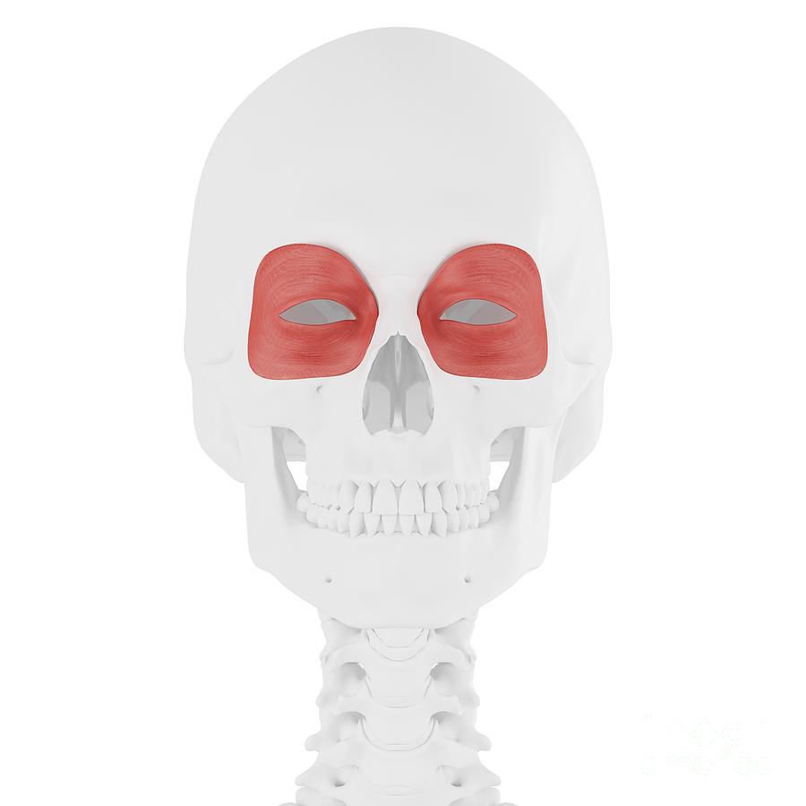
Orbicularis Oculi Muscle Twitching - LAB PRACTICAL LO'S (WEEK 2) flashcards | Quizlet, It is further divided into palpebral, lacrimal and ocular portions.. (3) a fibrous layer that gives the lid its mechanical stability, its principal portions being the tarsal plates, which border directly upon the opening between the lids, called the palpebral aperture; The orbicularis oculi is continuous with the superficial musculoaponeurotic system (smas) in the upper face as is the platysma in the lower. Your orbicularis oculi are muscles that surround your eyes and help open and close your eyelids. Joe muscolino october 17, 2017. It lies in the tissue of the eyelid and causes the eye to close or blink.
It can also contribute to twitching of the eye and a drooping eyelid. At the same time, it compresses the nearby tear gland, or lacrimal gland, aiding the flow of tears over the surface of the eye. Orbicularis oculi is a skeletal muscle of the face that surrounds the eye and is responsible for closing the eye. The orbicularis oculi (also orbicularis oculi muscle, latin: Train orbicularis oculi muscles, 3.
cheek raiser vs. lid tightener - Face the FACS from i0.wp.com Together with corrugator supercilii and orbicularis oculi consists of an orbital, palpebral and deep palpebral part. The orbicularis oculi is a muscle in the face that closes the eyelids. Musculus orbicularis oculi) is a circular shaped muscle around the opening of the eye. The main cause for overuse of this muscle is poor eyesight without. Blood supply of the orbicularis oculi is provided by branches of the facial, superficial temporal, maxillary and ophthalmic arteries. The muscle extends between three bones of the viscerocranium (frontal bone. At the same time, it compresses the nearby tear gland, or lacrimal gland, aiding the flow of tears over the surface of the eye. The muscle contributes to pain above the eye that travels down the side of the nose.
Questions and answers on orbicularis oculi muscle.
Anterior view of a myofascial trigger point and its referral zone in the orbicularis oculi, right side of the body. The main cause for overuse of this muscle is poor eyesight without. Gross anatomy the orbicularis oculi is subdivided into orbital, palpebral and lacrimal parts.each has defined actions. Velg blant mange lignende scener. Injury to the orbicularis oculi can result from overuse, which may result in headaches, eyestrain, or sinus headaches. Information on the orbicularis oculi by the anatomyzone daily feed. Orbicularis oculi is not attached to any bones on the lateral side that is why when you close your eyelids, they are both drawn to the middle 2. Your orbicularis oculi are muscles that surround your eyes and help open and close your eyelids. (3) a fibrous layer that gives the lid its mechanical stability, its principal portions being the tarsal plates, which border directly upon the opening between the lids, called the palpebral aperture; The muscle comprises the following three sections: The orbicularis oculi (also orbicularis oculi muscle, latin: My left upper eye lid has been twitching for 2 weeks. Together with corrugator supercilii and orbicularis oculi consists of an orbital, palpebral and deep palpebral part.
Orbicularis oculi muscle action buccinator muscle action orbicularis oculi muscle orbicularis oris action zygomaticus major and minor. It is further divided into palpebral, lacrimal and ocular portions. Orbicularis oculi muscle is generally a circular muscle located below your skin, around your eyes4. At the same time, it compresses the nearby tear gland, or lacrimal gland, aiding the flow of tears over the surface of the eye. Orbicularis oculi is a circular thin muscle.
Pin on Ideas from i.pinimg.com The main cause for overuse of this muscle is poor eyesight without. The orbicularis oculi muscle is a ringlike band of muscle, called a sphincter muscle, that surrounds the eye. Injury to the orbicularis oculi can result from overuse, which may result in headaches, eyestrain, or sinus headaches. The orbicularis oculi (also orbicularis oculi muscle, latin: The muscle extends between three bones of the viscerocranium (frontal bone. (3) a fibrous layer that gives the lid its mechanical stability, its principal portions being the tarsal plates, which border directly upon the opening between the lids, called the palpebral aperture; Orbicularis oculi definition it is a circular band of muscle that surrounds the eye. Blood supply of the orbicularis oculi is provided by branches of the facial, superficial temporal, maxillary and ophthalmic arteries.
Train orbicularis oculi muscles, 3.
It lies in the tissue of the eyelid and causes the eye to close or blink. The muscle extends between three bones of the viscerocranium (frontal bone. Musculus orbicularis oculi) is a circular shaped muscle around the opening of the eye. Part of the reason can be attributed to the complicated spatial arrangement of the myofibers and their insertion directly into the skin of the face. Orbicularis oculi muscle to supraorbital nerve stimulation: (3) a fibrous layer that gives the lid its mechanical stability, its principal portions being the tarsal plates, which border directly upon the opening between the lids, called the palpebral aperture; Gross anatomy the orbicularis oculi is subdivided into orbital, palpebral and lacrimal parts.each has defined actions. My left upper eye lid has been twitching for 2 weeks. This is training orbicularis oculi (facial muscle) by mike talani on vimeo, the home for high quality videos and the people who love them. It has skeletal muscles and nerves from the this part of orbicularis oculi is skeletal and pale in color and starts from the splitting of tendo oculi and traverses both of the eyelids to mix sideways with eye. It is subdivided into the pretarsal, preseptal, and orbital muscles. The orbicularis oculi muscle is the muscle that encircles the eye. It is generally a sphincter muscle that lies in the tissue of the orbicularis oculi description.
What causes twitching in left upper eyelid? Blood supply of the orbicularis oculi is provided by branches of the facial, superficial temporal, maxillary and ophthalmic arteries. It is subdivided into the pretarsal, preseptal, and orbital muscles. Anterior view of a myofascial trigger point and its referral zone in the orbicularis oculi, right side of the body. It surrounds the eye hence named so.
Orbicularis Oculi Muscle Photograph by Sebastian Kaulitzki ... from images.fineartamerica.com It has skeletal muscles and nerves from the this part of orbicularis oculi is skeletal and pale in color and starts from the splitting of tendo oculi and traverses both of the eyelids to mix sideways with eye. It is generally a sphincter muscle that lies in the tissue of the orbicularis oculi description. The orbicularis oculi is a muscle in the face that closes the eyelids. Joe muscolino october 17, 2017. Orbicularis oculi trigger point diagram, pain patterns and related medical symptoms. Oc orbicularis oculi muscle, or. It lies in the tissue of the eyelid and causes the eye to close or blink. It help in the passing and draining of tears.
Orbicularis oculi is not attached to any bones on the lateral side that is why when you close your eyelids, they are both drawn to the middle 2.
It can also contribute to twitching of the eye and a drooping eyelid. Gross anatomy the orbicularis oculi is subdivided into orbital, palpebral and lacrimal parts.each has defined actions. The orbicularis oculi muscle is a ringlike band of muscle, called a sphincter muscle, that surrounds the eye. The myofascial pain pattern has pain locations that are displayed in red and associated trigger points shown as xs. It lies in the tissue of the eyelid and causes the eye to close or blink. Your orbicularis oculi are muscles that surround your eyes and help open and close your eyelids. Orbicularis oculi is not attached to any bones on the lateral side that is why when you close your eyelids, they are both drawn to the middle 2. The orbicularis oculi is continuous with the superficial musculoaponeurotic system (smas) in the upper face as is the platysma in the lower. Try our newest study sets that focus on orbicularis oculi muscle action to increase your studying efficiency and retention. Musculus orbicularis oculi) is a circular shaped muscle around the opening of the eye. Orbicularis oculi trigger point diagram, pain patterns and related medical symptoms. Velg blant mange lignende scener. Injury to the orbicularis oculi can result from overuse, which may result in headaches, eyestrain, or sinus headaches.
…muscular layer containing principally the orbicularis oculi muscle, responsible for lid closure; orbicularis oculi muscle. Orbicularis oris muscle, stc sensory trigeminal complex, nts.


 flashcards | Quizlet, It is further divided into palpebral, lacrimal and ocular portions.)